- Informed consent, timeout.
- Perform screening U/S to ID landmarks and r/o significant vascular structures in field.
- Commence sedation (in addition to sedation plan, give strong consideration to short-acting neuromuscular paralytic such as rocuronium).
- Position with neck extended (with pillow or towels under shoulders), prep and drape (note: chlorhexidine is preferred to the iodide shown in the photo):

- Use sterile pen to mark thyroid cartilage, crycothyroid membrane, and the first several tracheal rings:

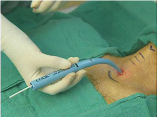
- Anesthetize with 1% lidocaine with epinephrine.
- Check the Shiley trach balloon, then hub the Shiley trach over the corresponding dilator (6 Shiley = 26 F dilator, 8 Shiley = 28 F dilator).
- Make horizontal 1.5-2 cm skin incision over space between 1st and 2nd tracheal rings or between 2nd and 3rd tracheal rings.

- Perform blunt dissection with forceps and gloved finger down to level of trachea:

- Second operator advances bronchoscope via swivel adapter to the just past the end of the ETT tube. Anteroflex the scope with the tip just past the end of the ETT.
- Deflate the ETT cuff and slowly pull back until transillumination can be seen under the incision:

- The gloved finger pushing on the anterior wall of the trachea should be seen in the scope view. Pull the ETT back 1 cm more and leave the bronchoscope just inside the end of the ETT, pointing down toward the center of the trachea.
- Advance a needle or angiocath with mounted syringe with saline inside into the trachea, trying to pass midway between the tracheal rings. Demonstrate return of air bubbles. Confirm position bronchoscopically:

- Confirm that the needle has not passed through the ETT by having the second operator or RT wiggle the ETT down and up slightly and show that this does not move the needle.
- Advance the green wire roughly to carina depth under bronchscopic guidance, then remove the needle or vascath:

- Perform initial dilatation with 14F dilator then remove off wire:

- Place guide catheter so that the safety ridge is aligned with the skin and the silve mark on the green wire align with the back of the guid catheter:


- Activate the coating on the Blue Rhino with saline, then place the Blue Rhino so that its tip aligns with the safety ridge on the guide catheter at the level of the skin:
- Move the entire setup forward in an arc motion with to and fro movements, pausing for several seconds at a few inches depth:

- Advance until the resistance is overcome and align the black mark on the Blue Rhino with the skin. Leave in place a few seconds:

- Bring the Blue Rhino and guide catheter out until the safety ridge on the guide catheter is back at skin level, then remove the Blue Rhino off the guide catheter.
- Lubricate the Shiley/dilator combination with surgilube, then place the tip at the safety ridge on the guide catheter, then advance the whole setup until the trach is in place:


- Remove the dilator, guide catheter, and wire, inflate trach balloon.
- Place the inner cannula and attach the ventilator tubing.

- Confirm position by bronchoscopy and return of appropriate ventilator volumes, bilateral breath sounds.
- Secure in position at the four corners with sutures, then apply a Shiley velcro trach tie:

Full procedure video on Cook site:
http://www.cookmedical.com/cc/datasheetMedia.do?mediaId=1557&id=4011
Important Disclaimer: No warranty whatsoever is made that any of the information contained herein is accurate. There is absolutely no assurance that any statement in any posting on neurocriticalcare.pbwiki.com is true, correct, precise, or up-to-date. The medical information provided on neurocriticalcare.pbwiki.com cannot substitute for clinical judgment, training, and experience. The information on neurocriticalcare.pbwiki.com should never serve as a substitute for the advice of a physician trained in the care of critically ill patients with neurological or neurosurgical disease. None of the contributors to neurocriticalcare.pbwiki.com take any responsibility for the results or consequences of any attempt to use or adopt any of the information presented in any of the postings to this web site. None of the postings to neurocriticalcare.pbwiki.com should be construed as an attempt to offer or render a medical opinion or otherwise engage in the practice of medicine. The postings on neurocriticalcare.pbwiki.com may be modified by several different individuals, and therefore the postings do not represent the views or advice of any individual, institution, or entity.
Comments (0)
You don't have permission to comment on this page.